Fitxategi:Free-living amebic infections.png
Orriaren edukia ez da onartzen beste hizkuntza batzuetan.
Wikipedia, Entziklopedia askea

Bereizmen handikoa((1.365 × 1.170 pixel, fitxategiaren tamaina: 715 KB, MIME mota: image/png))

Fitxategi hau Wikimedia Commonsekoa da. Hango deskribapen orriko informazioa behean duzu.
Commons gordailu bat da, lizentzia askea duten multimedia fitxategiena. Lagun dezakezu.

Fitxategi hau Wikimedia Commonsekoa da
Irudi hau birsortu beharko litzateke SVG irudi bektorial moduan. Honek abantaila asko ematen ditu, ikus Commons:Media for cleanup (ingelesez) informazio gehiagorako. Irudi honen SVG bertsioa eskuragarri badago, mesedez Commonsera igo. Igo ostean, ordezkatu txantiloi hau txantiloi honegatik {{vector version available|irudiaren izen berria.svg}} irudi honetan.

Laburpena

Deskribapena
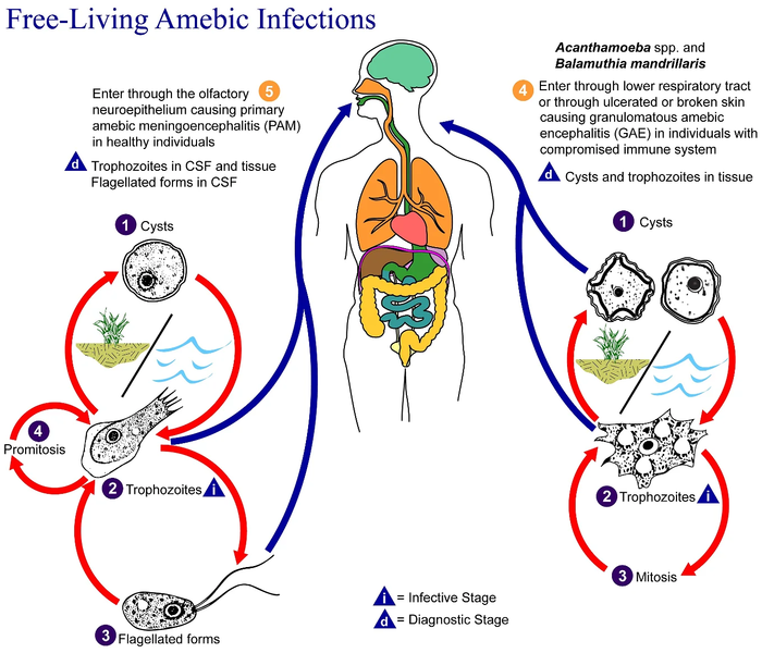
English: This is an illustration of the life cycle of the parasitic agents responsible for causing “free-living” amebic infections.

For a complete description of the life cycle of these parasites, select the link below the image or paste the following address in your address bar: http://www.dpd.cdc.gov/dpdx/HTML/FreeLivingAmebic.htm Free-living amebae belonging to the genera Acanthamoeba, Balamuthia, and Naegleria are important causes of disease in humans and animals. Naegleria fowleri produces an acute, and usually lethal, central nervous system (CNS) disease called primary amebic meningoencephalitis (PAM). N. fowleri has three stages, cysts (1) , trophozoites (2) , and flagellated forms (3) , in its life cycle. The trophozoites replicate by promitosis (nuclear membrane remains intact) (4) . Naegleria fowleri is found in fresh water, soil, thermal discharges of power plants, heated swimming pools, hydrotherapy and medicinal pools, aquariums, and sewage. Trophozoites can turn into temporary flagellated forms which usually revert back to the trophozoite stage. Trophozoites infect humans or animals by entering the olfactory neuroepithelium (5) and reaching the brain. N. fowleri trophozoites are found in cerebrospinal fluid (CSF) and tissue, while flagellated forms are found in CSF.

Acanthamoeba spp. and Balamuthia mandrillaris are opportunistic free-living amebae capable of causing granulomatous amebic encephalitis (GAE) in individuals with compromised immune systems. Acanthamoeba spp. have been found in soil; fresh, brackish, and sea water; sewage; swimming pools; contact lens equipment; medicinal pools; dental treatment units; dialysis machines; heating, ventilating, and air conditioning systems; mammalian cell cultures; vegetables; human nostrils and throats; and human and animal brain, skin, and lung tissues. B. mandrillaris however, has not been isolated from the environment but has been isolated from autopsy specimens of infected humans and animals. Unlike N. fowleri, Acanthamoeba and Balamuthia have only two stages, cysts (1) and trophozoites (2) , in their life cycle. No flagellated stage exists as part of the life cycle. The trophozoites replicate by mitosis (nuclear membrane does not remain intact) (3) . The trophozoites are the infective forms and are believed to gain entry into the body through the lower respiratory tract, ulcerated or broken skin and invade the central nervous system by hematogenous dissemination (4). Acanthamoeba spp. and Balamuthia mandrillaris cysts and trophozoites are found in tissue.
Data
Jatorria

This media comes from the Centers for Disease Control and Prevention's Public Health Image Library (PHIL), with identification number #3412.

Note: Not all PHIL images are public domain; be sure to check copyright status and credit authors and content providers.


العربية | Deutsch | English | македонски | slovenščina | +/−

Egilea
  • Photo Credit:
  • Content Providers(s): CDC/Alexander J. da Silva, PhD/Melanie Moser
Lizentzia
(Fitxategi hau berrerabiltzen)
English: None - This image is in the public domain and thus free of any copyright restrictions. As a matter of courtesy we request that the content provider be credited and notified in any public or private usage of this image.
Beste bertsioak

Lizentzia

Public domain
This image is a work of the Centers for Disease Control and Prevention, part of the United States Department of Health and Human Services, taken or made as part of an employee's official duties. As a work of the U.S. federal government, the image is in the public domain.

eesti  Deutsch  čeština  español  português  English  français  Nederlands  polski  slovenščina  suomi  македонски  українська  日本語  中文(简体)‎  中文(繁體)‎  العربية  +/−

Jatorrizko igoera erregistroa

Transferred from en.wikipedia to Commons by Optigan13.

The original description page was here. All following user names refer to en.wikipedia.
  • 2006-05-04 01:09 Keenan Pepper 518×435×8 (31658 bytes) Free-living_amebic_infections.gif, converted to [[PNG]] by [[netpbm]].

Irudi-oineko testuak

Add a one-line explanation of what this file represents

Fitxategi honetan agertzen diren itemak

honako hau irudikatzen du

MIME type ingelesa

image/png

Fitxategiaren historia

Data/orduan klik egin fitxategiak orduan zuen itxura ikusteko.

Data/OrduaIruditxoaNeurriakErabiltzaileaIruzkina
oraingoa11:24, 2 otsaila 202311:24, 2 otsaila 2023 bertsioaren iruditxoa1.365 × 1.170 (715 KB)Materialscientisthttps://answersingenesis.org/biology/microbiology/the-genesis-of-brain-eating-amoeba/
08:30, 20 uztaila 200808:30, 20 uztaila 2008 bertsioaren iruditxoa518 × 435 (31 KB)Optigan13{{Information |Description={{en|This is an illustration of the life cycle of the parasitic agents responsible for causing “free-living” amebic infections. For a complete description of the life cycle of these parasites, select the link below the image

Ez dago fitxategi hau darabilen orririk.

Fitxategiaren erabilera orokorra

Hurrengo beste wikiek fitxategi hau darabilte:

Metadatuak